Although it is one of the oldest paraclinic exams, dating back to the late 19th century, the ECG is still of crucial clinical use. This examination often still poses problems of interpretation to the medical practitioner.
This course aims to help the student, the practicing physician and even the trained cardiologist to improve his knowledge in electrocardiography. It consists of 250 traces of varying complexity with a description of each one by experts. This allows the reader to compare his analysis with that of the experts. In addition, the areas of interest of the ECG can be activated to be clearly highlighted.
We hope that these plots will be useful to readers and will improve their knowledge.
The ECGs are available sorted by keywords and categories.
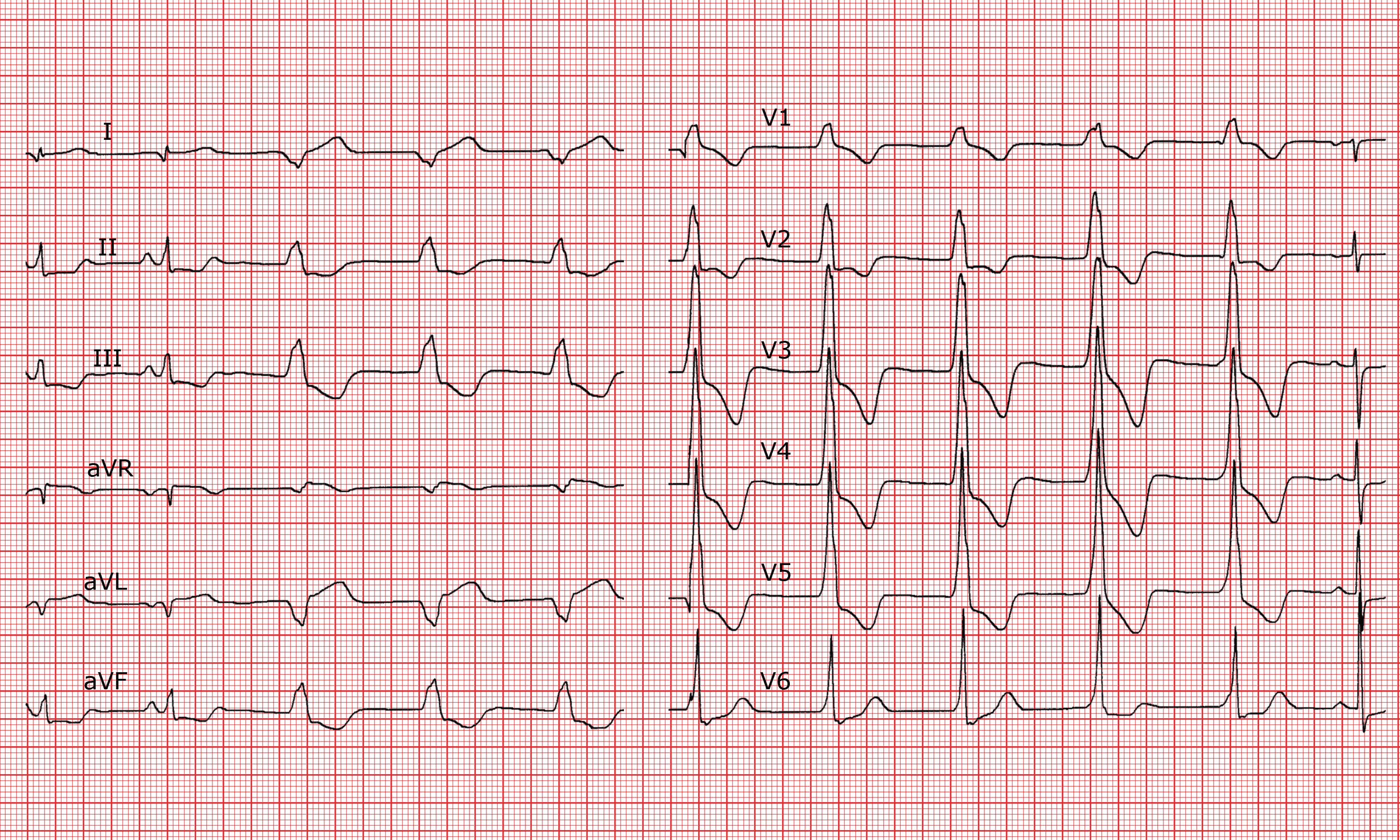
2 visible waves (second complex in the peripheral leads and last complex in the precordial leads).
PR interval
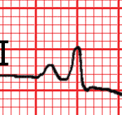
Normal.
QRS
For the 2 complexes in sinus rhythm there is a micropotential with a qr pattern in I and qs pattern in aVL. With the wide QRS complexes there is right axis deviation and an exclusive R wave in all precordial leads.
ST segment
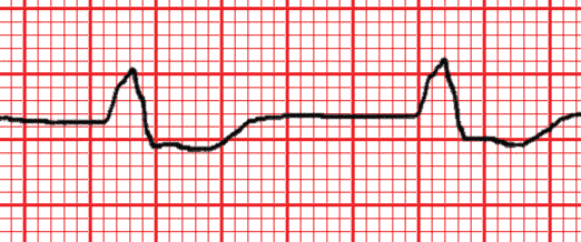
Elevation in I and aVL, depression in II, III and aVF for the narrow QRS complexes.
T waves
Negative in III for the narrow QRS complexes (diffuse changes inthe terminal phase of the wide QRS complexes).
QT interval
Normal.
Zones
Regular normal sinus rhythm.Wide QRS complexes regular rhythm.
Diagnostic
Accelerated idioventricular rhythm (AIVR) related to a lateral infarction.
Comments
The second narrow QRS complex in the peripheral leads and the last QRS complex in the precordial leads are normally preceded by a P wave; the basic rhythm is sinus rhythm. The subsequent QRS complexes are wide with P wave dissociation; the rhythm is regular, slightly faster than the baseline rhythm; this is sometimes called "slow ventricular tachycardia" but is better referred to as accelerated idioventricular rhythm (AIVR).
This arrhythmia generally arises during the coronary reperfusion phase after an infarction. In the present case it is shown by the Q waves in I and aVL and the ST segment depression in the inferior leads when there is a sinus rhythm.
The axis of the wide QRS complexes is deviated to the right with an R wave in V1, which allows us to locate the origin of the idioventricular rhythm in the proximity of the infarcted region.